Introduction
It is a classical approach to the lumbar spine for the fusion between 2 lumbar vertebrae from the back approach. This basic principle is the same as all different kinds of technique for spinal fusion. The commonest indications include disc degeneration with persistent back pain, acute disc hernia with unstable spinal segments, spinal stenosis and spondylolisthesis. This surgery is proven to be safe and effective in managing the lumbar disc problem with / without instability. All the spinal fusion surgeries are considered as major procedure due to its high demand of surgical skills and experience. The cost involved for this surgery is usually higher than other orthopaedic procedures.


Before the surgery, your surgeon will assess your condition that you are suitable for this procedure. After clinical examinations, X-rays include flexion extension view of the lumbar spine and MRI scan of the lumbar spine are usually enough for the decision. Once the surgery plan fixed, you should contact your insurance company for coverage of the surgery cost.


The operation usually takes 3.5 to 4 hours to complete. Normally, you can start mobilisation on the next day and stay in hospital for 4-5 days more for physiotherapy rehabilitation exercises. Normally, you can be discharged from hospital on day 5-7 after the surgery.


After some blood work and pre-anaesthestic assessment by our anaesthetist, consent forms are signed before the nurse can bring you to the operating room.
You will be under general anaesthesia with a tube inside your airway for ventilation. You will be positioned with your back facing up. Your face will be supported by a specially designed foam and the pressure points of the body will be padded. This procedure is usually required 1 surgeon and 1 assistant. The steps are complicated and time consuming.
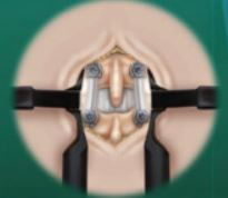
After draping with sterile drapes, the surgical area will be ready to start the procedure. The incision will be guided by intra-operative fluoroscopy. A midline incision will be made from one spinous process to the other (Fig.1).

Fig. 1 Exposure of the spine for the MidLIF - a modified posterior lumbar interbody fusion technique with least soft tissue trauma.
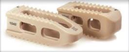
Minimal dissection of the back muscle from midline is required to gain the exposure of the posterior aspect of the spine. With a careful dissection, it will usually leave minimal trauma to the soft tissue around with the new technique. A special retractor will be used to keep the space for working. The screw entry site will be marked and confirmed with fluoroscopy before a laminectomy done. The bone from the laminectomy will be used as a bone graft for fusion inside the disc space. A plastic rectangular implant called inter body fusion cage (Fig. 2) will be inserted into the disc space. Standard PLIF surgery will require 2 cages, one on each side of the disc to provide better stability. The plastic cage is hollow in the middle where the bone graft can be packed in.

Fig. 2 Specially designed PLIF cages for lumbar spine fusion. They are hollow in the middle for placing bone graft for bone union.
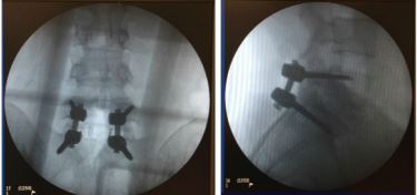
The fusion area is larger than with only one cage. Finally, pedicle screws are inserted at the lumbar spine, 2 screws at each level (total 3 screws will be used for 1 disc level fusion) to complete the procedure. Intra-operative fluoroscopy screening confirms all the implant position (Fig. 3).

Fig. 3 Intra-operative Fluoroscopy of the spine to confirm the implant position after the fusion.
The exposed nerve root and dura sac will be covered with anti-adhesion substance. Layers of tissues are repaired with strong sutures. A plastic tube (aka drain) will be placed just beneath the skin incision to drain the residual exudate or blood. The drain will usually be removed on day 1 or 2 after the surgery. The incisions are closed with absorbable sutures and finally covered with a layer of glue called Dermabond. Plastic waterproof dressing is covering the incision and the glue. You will be flip over to sleep on your back before the anaesthetist wake you up.


After the surgery, you will be placed at the recovery bay in the operating room for further monitoring. If there is not much wound pain or breathing difficulty, you will be returned to the ward for rest.
Theoretically, you can resume eating and drinking once you recovered from the anaesthesia. However, most of the patient with a few hours of anaesthesia, they will not have very good appetite to eat. We recommend to try some fluid, soup or congee on the first day. On the next day, you can resume your normal meal if the appetite is back.
Oral medications will be started on day of the surgery for pain management. The common side effects of the pain medicine are dizziness, nausea and vomiting. If you develop these side effects, please notify your nurse to take further action.
On day 1 after surgery, we will see your condition in the morning and physiotherapy will visit you shortly after. A soft lumbar corset will be applied and start some gentle exercises. You can start walking exercise and go to bathroom if the back wound pain allowed. There will not be any concern on the stability of the implant in usual activities. The drain on the back will be removed if there is not much output.
Day 2 to 5 will be the rehabilitation phase mainly taking care of the surgical wound pain and some walking exercises. You may experience the pain is different from the usual back pain. The leg numbness or sciatica pain may also gone immediately, or couple of weeks later. You can leave the hospital to go home when you can walk to the front door of the hospital.


After discharged from hospital, you can take any ground transportation (car or train) to return home. If you are coming from Shanghai or Beijing, you can take Ordinary commercial flight at Business class seat to return home. Luggage assistant and pre-arranged ground transportation is highly recommended. Appropriate documents can be provided to the airline if needed.


The sutures are all inside the incision and dissolve by itself in 3 months time. The glue covering the incision serving a protection layer to prevent the fluid go in or come out. You can treat it as a scalp of the a healing wound. The plastic dressing on the surface is to protect the glue from moisture. The glue can dissolve into water and fall off prematurely. You will need to change the dressing only if there is discharge from the wound, the edges peel off or water going in. Otherwise, we suggest you to keep the dressing intact as much as possible. It remains sterile if you have not open it. You should keep the dressing for 3 weeks when the wound should be complete healed. You just remove the dressing and expose the glue. The glue will fall off together with the skin debris while you shower. Do not peel off the glue yourself as there will be some raw area expose causing infection. You might be prescribed oral antibiotics if you have a higher risk of infection. You are highly recommended to finish the course of the antibiotic if there is no obvious side effects seen.


Our nurse will contact you for the follow up arrangement. Typically, the first follow up will be arranged on 2 weeks after the surgery mainly aimed for wound checking. X-ray lumbar spine will be required from time to time during the follow up. Further follow up at 6 weeks, 12 weeks, 6 months and 12 months will be required to monitor the healing of the fusion site. Bone healing usually take 3-6 months and there will be a radiological delay of the sign in bone healing shown on X-ray. L5S1 disc will usually overlap with the iliac crest and sometimes CT scan lumbar spine is required to confirm the healing.


The general anaesthetic risk will be explained by our anaesthetist while signing the consent with you. Generally, the anaesthetic risks are drug reaction, airway problem, heart attack, stroke, loosening of teeth, transient blindness (due to prone position), deep vein thrombosis, pneumonia etc.
For the general surgical risks, wound infection, bleeding, painful or hypertrophic scar are less than 3%. The usual blood loss is around 100-200 cc, depends on how complex is the pathology. The neurological risk such as nerve root injury, dura sac tear required repair and superficial nerve (lateral cutaneous nerve of thigh) injury are possible but rare. Death and paralysis is extremely rare. For single level fusion, the implant infection rate is around 1%. The fusion rate is usually quoted as 95% by using rBMP mixed with autograft. The symptomatic non-union requiring revision is around 0.5%. In our experience, we have not encountered any deep wound infection in the last 5 years. Revision surgery for non-union of the fusion is around 3 cases in the last 5 years.


It is considered as one of the definitive surgical treatment option for discogenic back pain. If you have gone through a comprehensive nonsurgical treatment for more than 6 months with suboptimal result, you can consider this as the next step. The fusion is a long term solution for painful disc and there will not be any change of the segment you have fused. However, the remaining unfused segments will continue to have degeneration and causing problem in the future.


There are some concerns on the stress migrated to the disc above and below the level you have surgery. Some scientist postulated that the stress will accelerate the speed of the degeneration and leading to another surgery. However, up to this moment, there is no evidence showing this postulation is true and with high incidence. All the available evidence can only showed some degeneration might happen in a patient with fusion surgery done. With the modification of the current technique, we believe the risk of the damage to the adjacent segment structure will be much less and indirectly reducing the risk on this. Actually, even without the fusion, the adjacent segment may also degeneration and cause the problem again. We believe, if there is indication of a fusion surgery, this is not considered as a true risk.


The Posterior lumbar interbody fusion is one of a surgical option for back pain or sciatic pain management. It is safe, effective and reliable for spinal problem. Long term complications are minimal and it can be done with less invasive approach. Please contact our office if you have any further questions not included in this document.
All Rights Reserved. Web Design by YSD


